
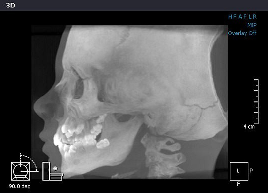

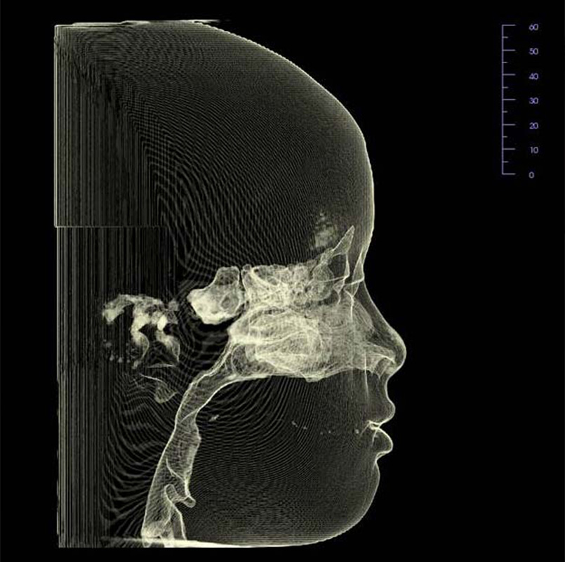
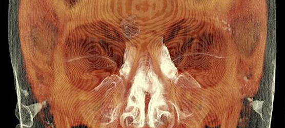
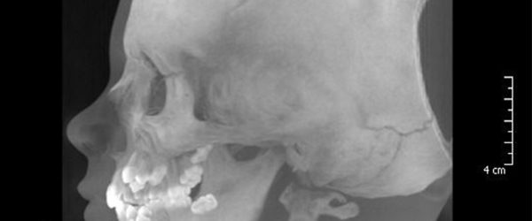
CBCT is a digital x-ray scanner designed for scanning the face, teeth and jaws. The scanner rotates 360° around a patient’s head in just seconds. The cone shaped x-ray beam provides data that can be formatted to produce 3D volume images for advanced planning and diagnostic support.
Orbit Imaging produces several Report Types and Conversions & Models suited for implantology, periodontics, orthodontics and endodontics. The superior 3D imaging technology includes DICOM file, Image Report, Viewer, and all digital images and diagnostic reports may be downloaded from ORBIT’s secure servers.
CBCT REPORTS, CONVERSIONS & MODELS
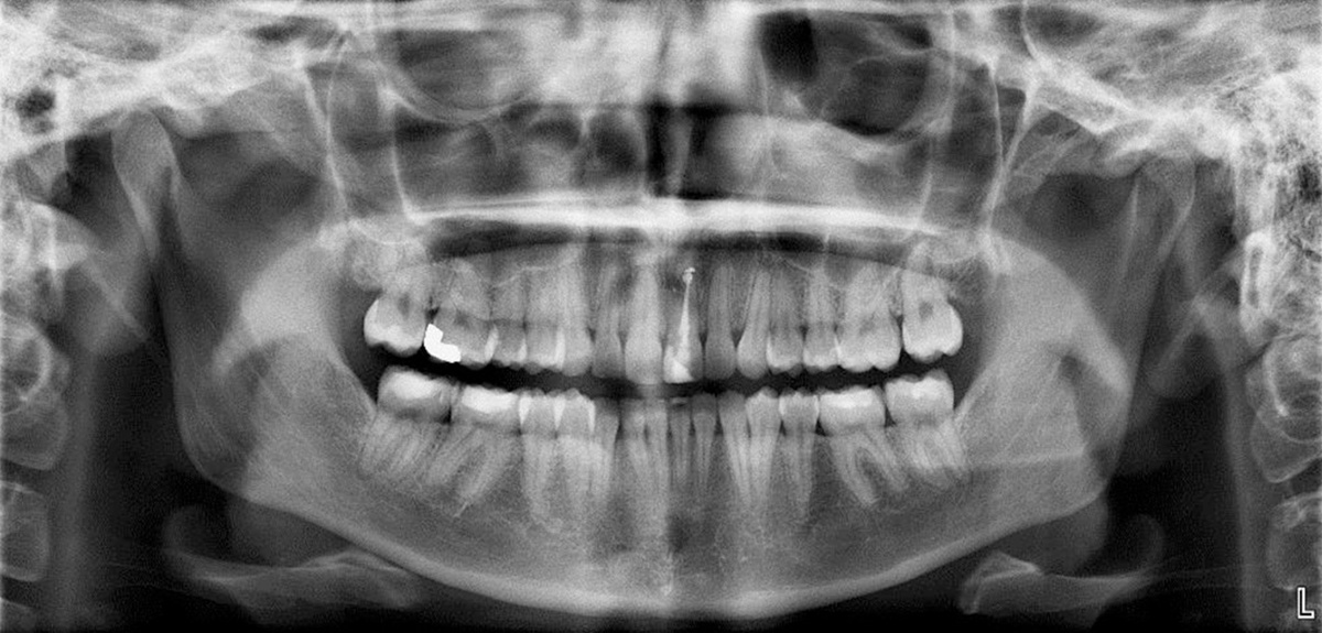
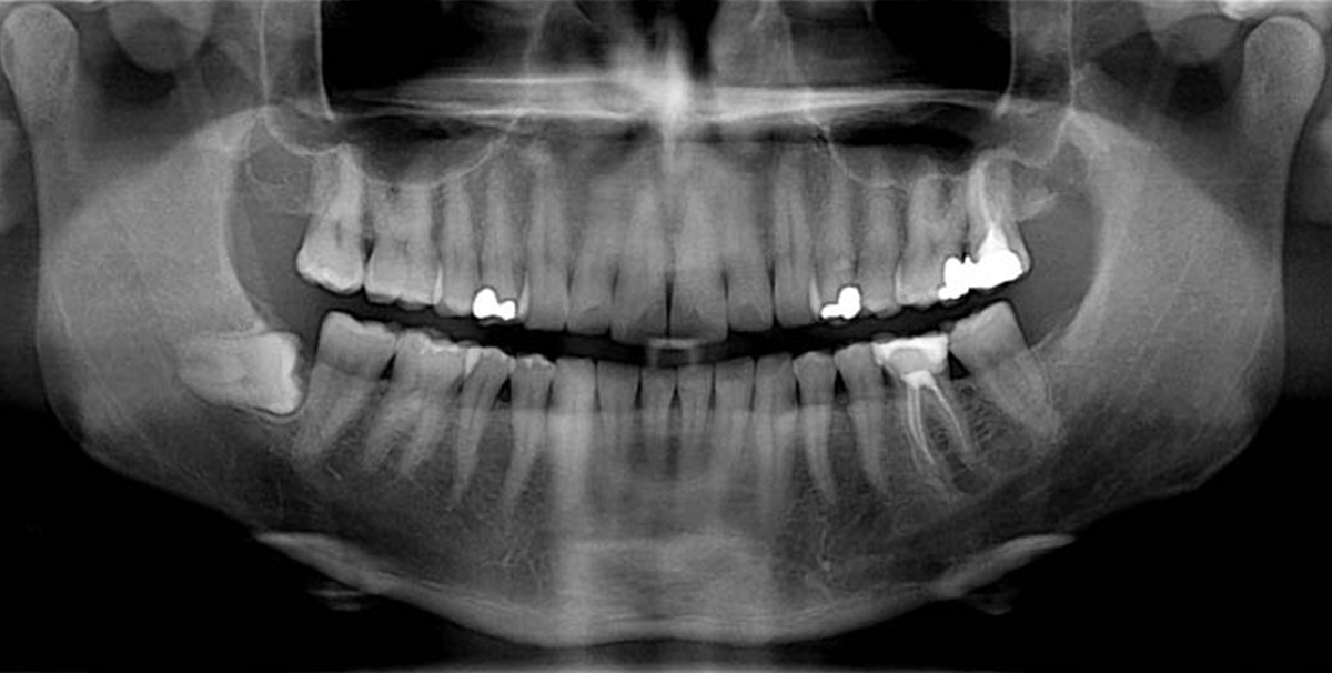
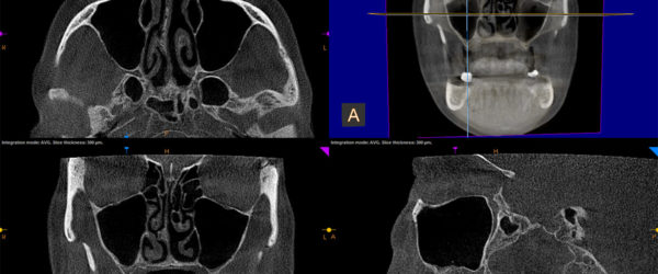
Orbit minimizes scanning time and reduces patient exposure while obtaining DICOM compliant volumetric views of all anatomic structures for bone and sinus assessment for dental implants, evaluation of impacted teeth and wisdom teeth, and investigation of airway, jaw and sinus pain and/or trauma.
CBCT imaging is available for single jaw, dual jaw and extended view imaging. Dual scans include a separate DICOM file of scan appliance.
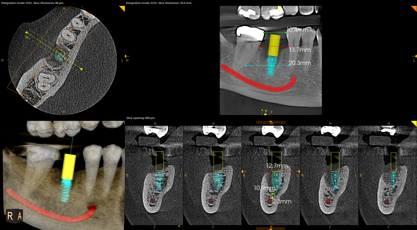
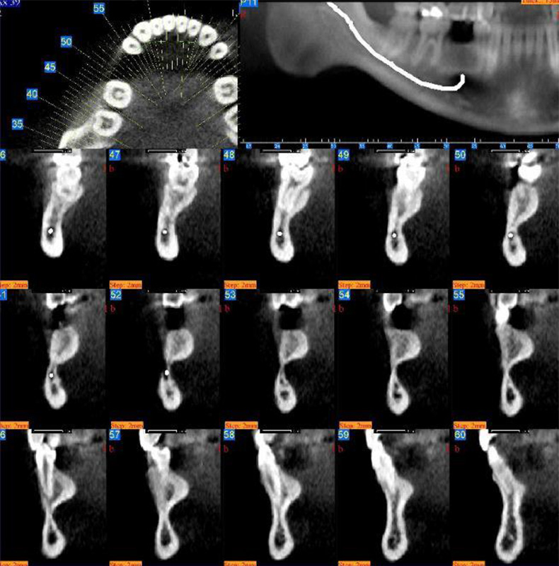
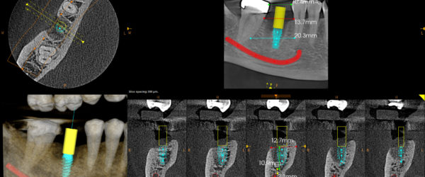
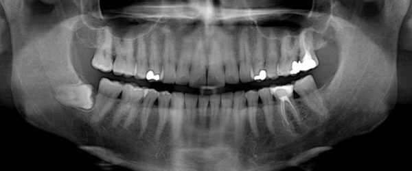
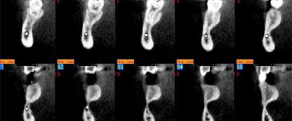
- Implant – A region of interest can be a localized site or the entire mandibular or maxillary arch. It consists of cross sections and panoramic projections and annotation of the nerve canal. It includes critical measurements of bone for optimum implant placement and 3D volumetric reconstruction of important anatomy. Conversion to Simplant and Nobel Guide useable formats, Implant planning and surgical guides are also available. Report is beneficial for diagnosis and treatment planning of implant sites and case presentation to the patient.
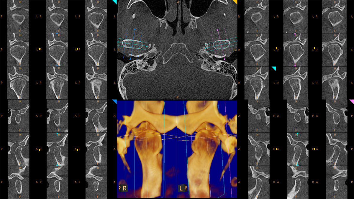
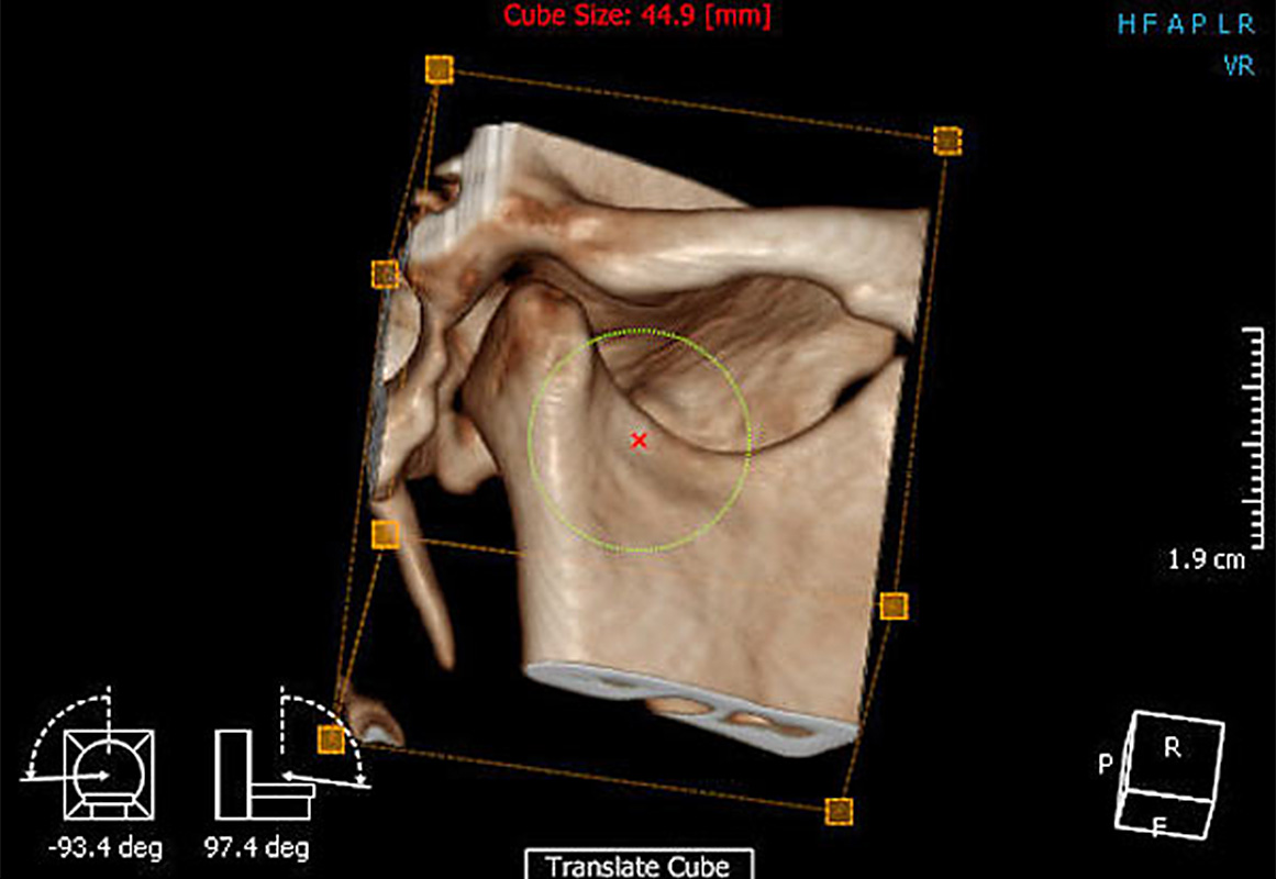
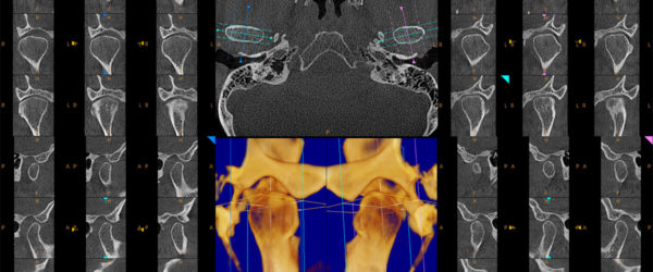
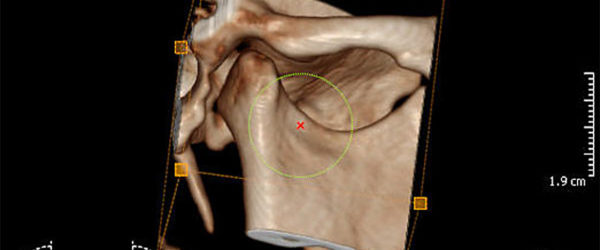
- Temporomandibular joint (TMJ) dysfunction (TMD) – TMJ Survey includes lateral, coronal and axial projections of the TMJ as well as panoramic and volumetric views.
- Surgical Planning
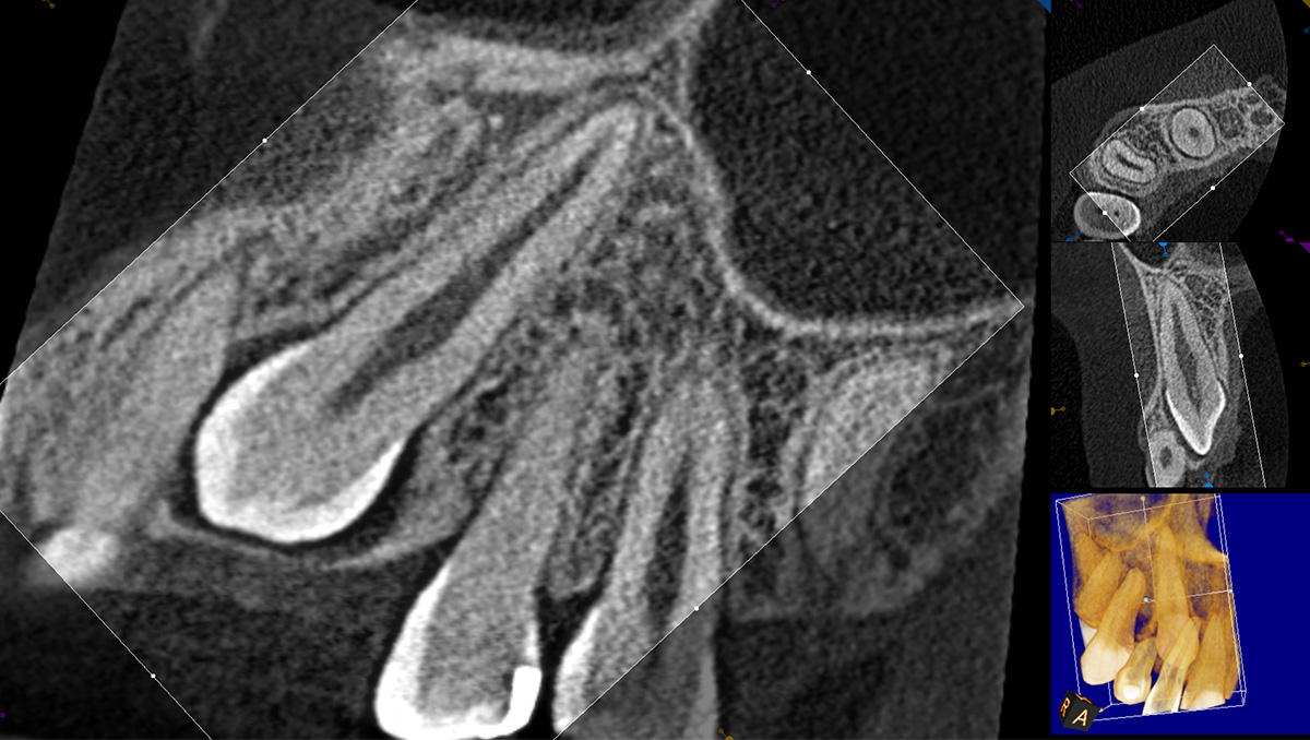
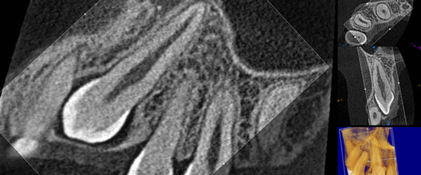
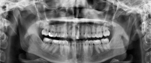
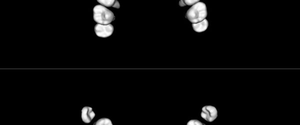
- 3rd Molars
- Impactions
- Supernumerary Teeth
- Pathology
- Cysts
- Tumors
- Foreign Bodies
- Orthodontic
- Anatamodel
- 3D Virtual Study Models
- Cephalometric Analysis
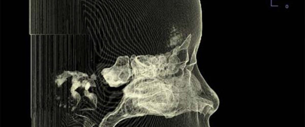
- Oral-Nasal Airways
- Sleep Apnea Diagnosis
- Trauma/Pathology
- Radiology Report – Scans and images can be reviewed by a board certified Oral & Maxillofacial Radiologist and a written diagnostic interpretive report generated on request to aid in patient diagnosis.
- Nobel Guide & Simplant Conversion – High quality conversion of DICOM image data from Cone Beam Volumetric Imaging and MDCT into SimPlant® (Planner, Pro and Master) & Facilitate™ readable formats.
- Implant Planning – Orbit technicians can convert your DICOM data to Simplant and Nobel Guide friendly formats, help you plan your case or provide you with a surgical guide.