
Imaging & Reporting Services
Refer patients for imaging or upload your existing CBCT studies for specialist reporting.
Patient Imaging
Patient imaging tailored to support diagnosis, treatment planning and image-guided care.
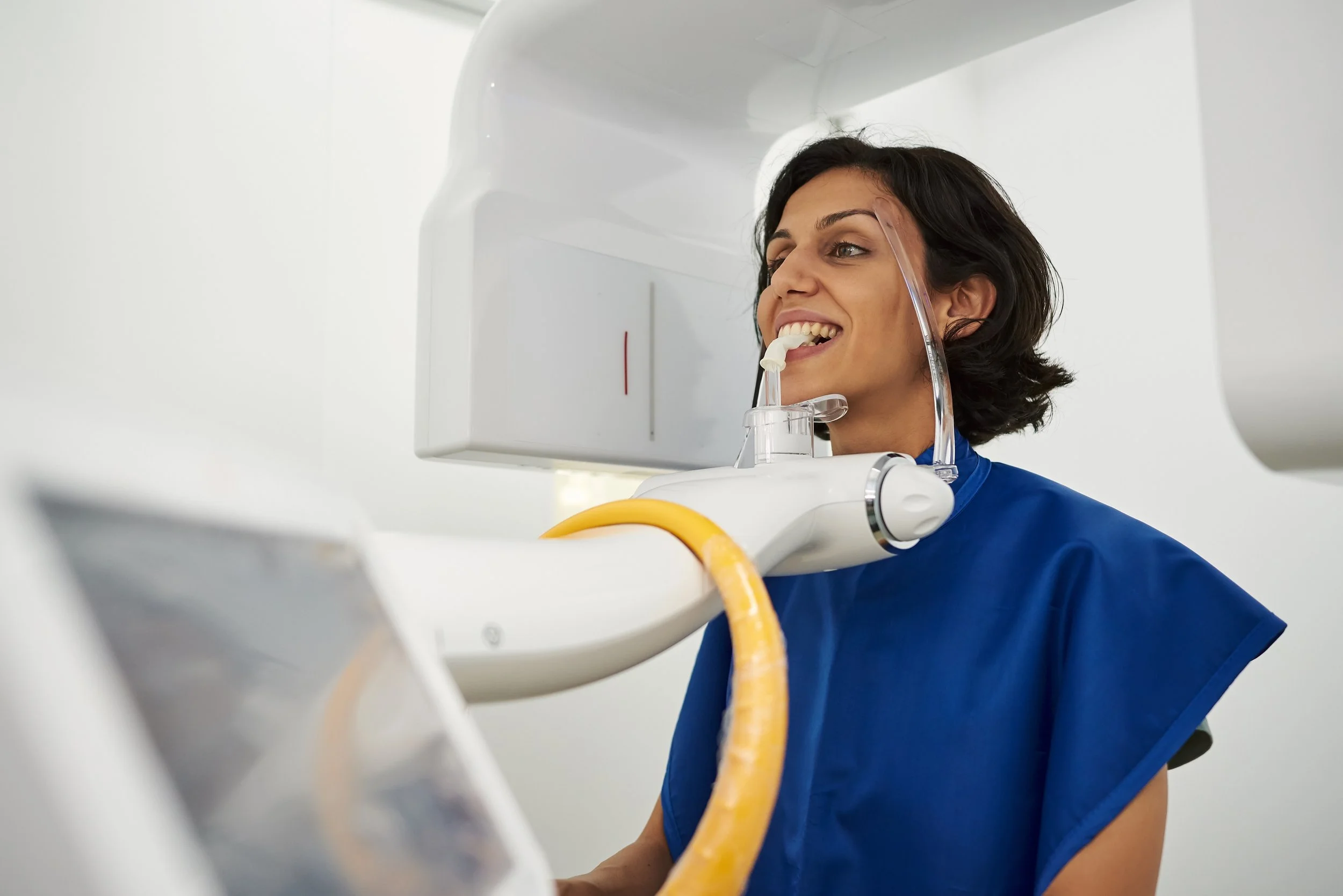
Cone Beam CT
Low-dose CBCT imaging with selectable fields of view from focused 3–4 tooth regions to extended cephalometric acquisition. Supports implant planning, endodontics, orthodontics, TMJ assessment, oral surgery and other diagnostic applications.
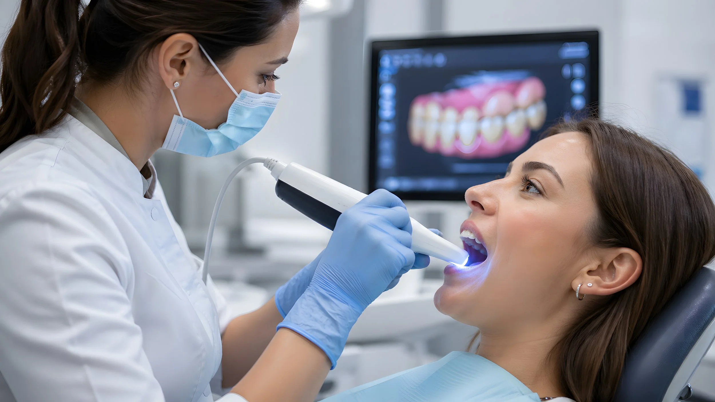
Intraoral Scanning (IOS)
High-resolution digital impressions for restorative, orthodontic and treatment-planning workflows. STL files can be provided for direct integration with your preferred laboratory, aligner or digital workflow.
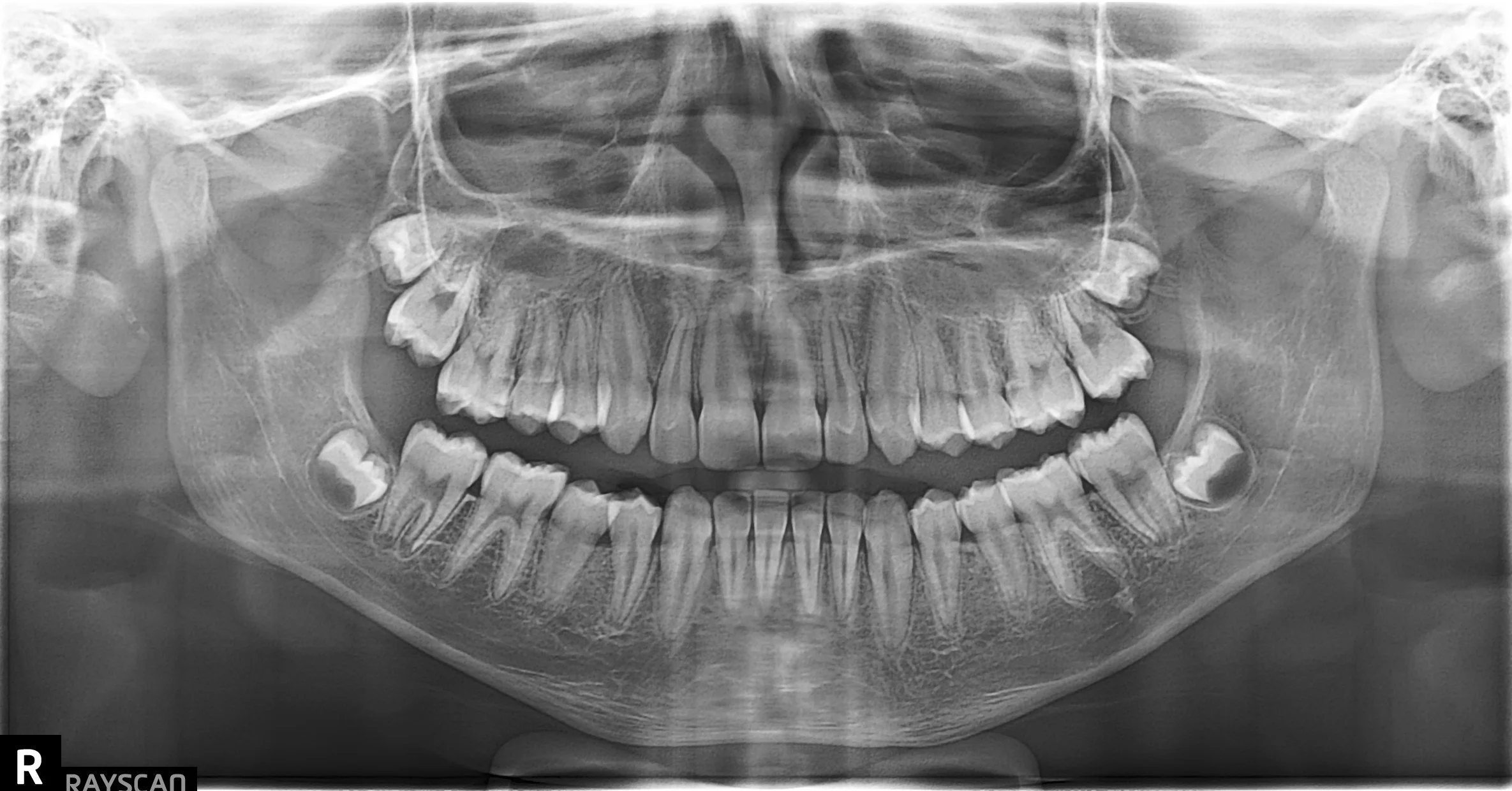
Panoramic
High-quality panoramic imaging providing a broad overview of the teeth, jaws, TMJs and surrounding structures for general diagnostic and treatment-planning applications.
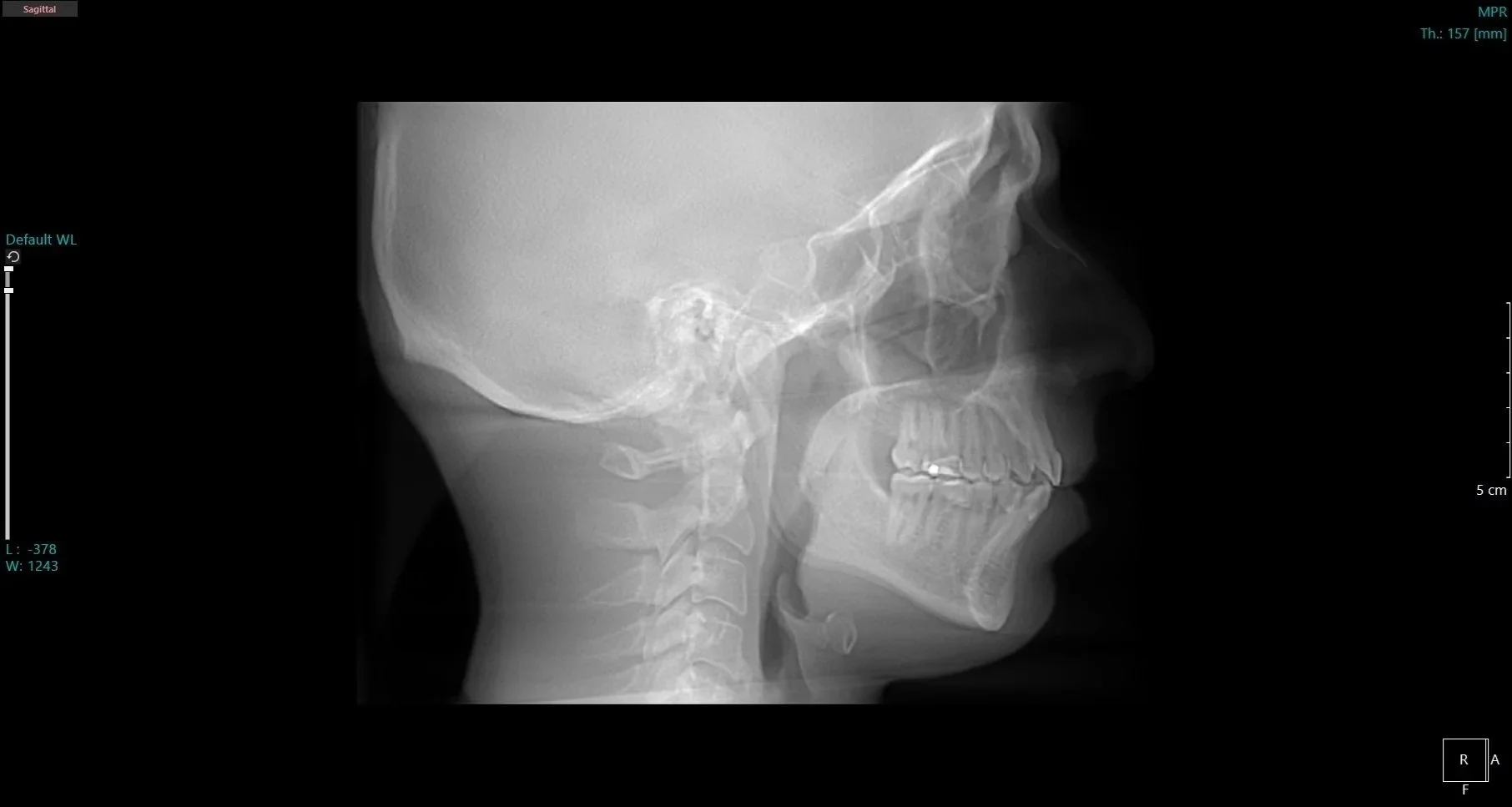
Cephalometric
Lateral and frontal cephalometric imaging for orthodontic and maxillofacial diagnosis and treatment planning. Cephalometric tracing and analysis are also available when requested.
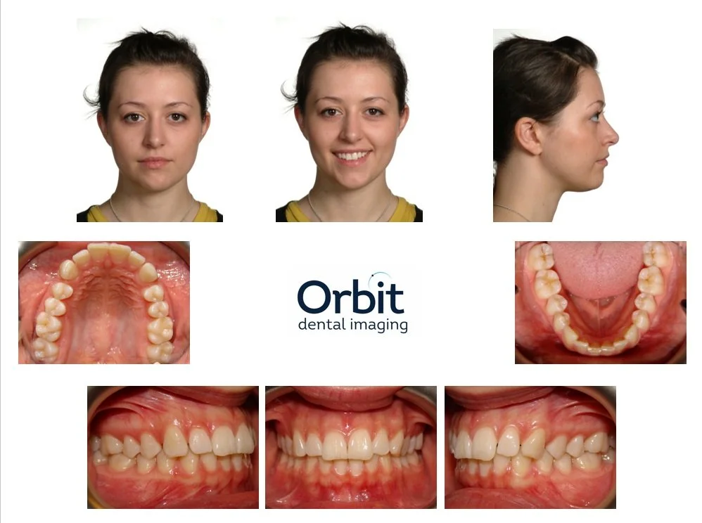
Clinical Photography
Standardized extraoral and intraoral clinical photography to support treatment planning, case documentation, interdisciplinary communication and patient consultation.
Radiology & Image Reporting
Specialist interpretation and reporting that transform your imaging data into clinically relevant information to support diagnosis, treatment planning and patient communication.
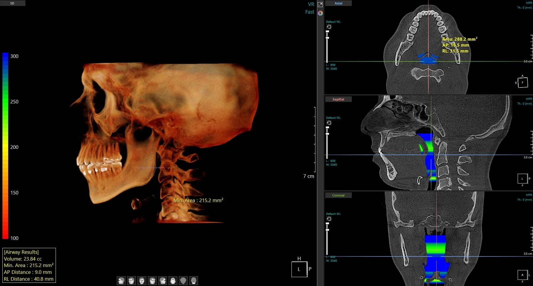
Radiology Reports
Specialist interpretation of the complete CBCT volume by Board Certified Oral Maxillofacial Radiologists, documenting relevant findings and impressions.
Are focused on your clinical question while providing review of the full imaged volume, with supporting images highlighting relevant anatomy and areas of interest.
Provide clear, actionable reporting with recommendations for follow-up where appropriate and direct access to the radiologist for case-related questions when needed.
Standard turnaround of 3-5 business days.
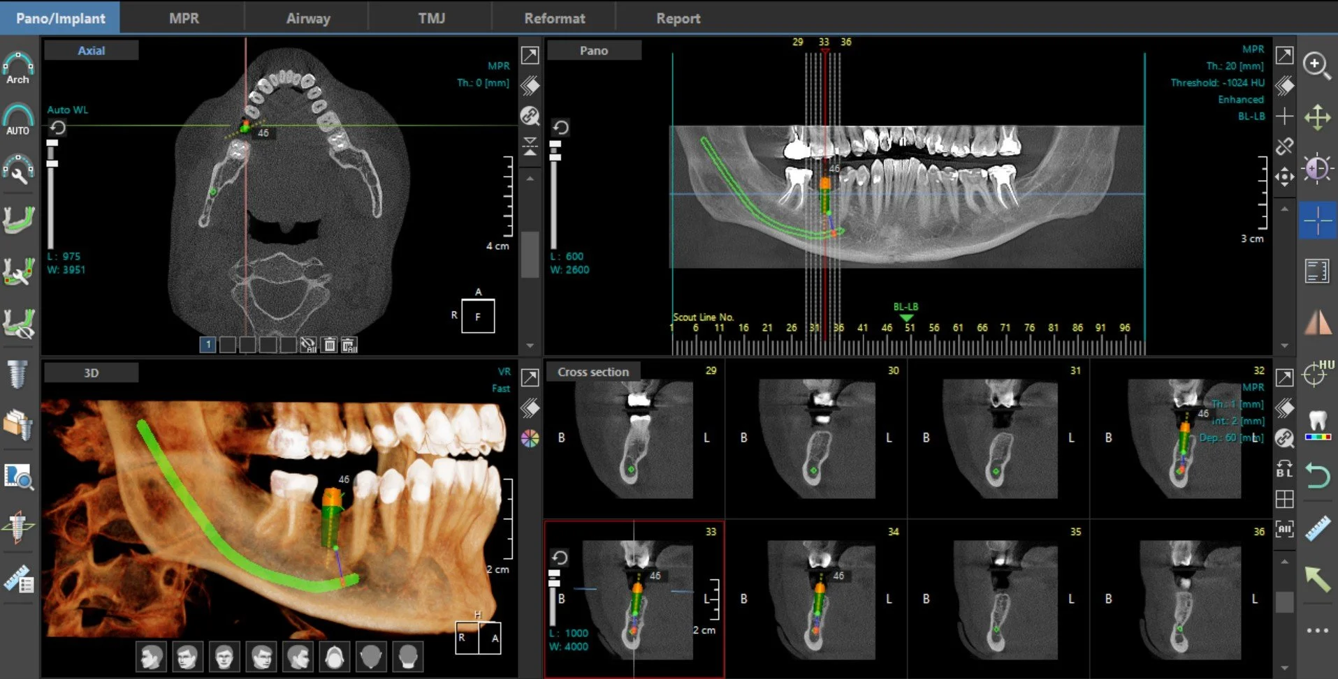
Image Portfolios
Customized views, reformats, renderings and measurements organized around the specific clinical application to support treatment planning and communication.
Relevant anatomy is clearly presented using multiplanar, cross-sectional, 3D and enhanced reconstructed views as appropriate to the case.
Measurements and calibrated imaging tools provide additional information for treatment planning when required.
Designed for practical clinical use, including treatment planning, case documentation and communication with patients and colleagues.
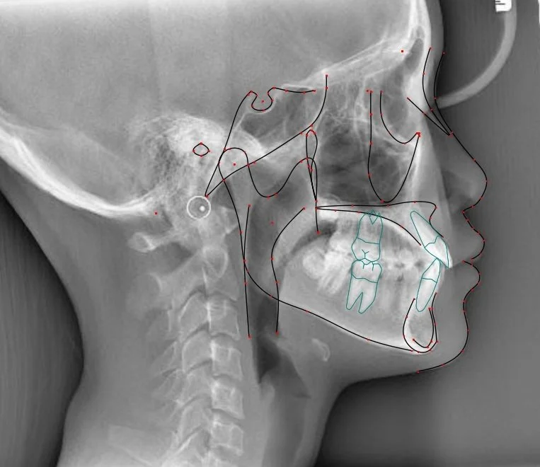
Cephalometric Analysis
2D & 3D cephalometric tracing and analysis using more than 90 available landmarks, with standard or customized analyses for orthodontic and orthognathic and maxillofacial treatment planning.